
Brain disease is diagnosed with various names and stages of development. Some of the most prevalent brain diseases are infections, tumours, epilepsy, trauma and stroke. The key to either conquering or containing most diseases is early detection.
Technology is at the forefront of early detection for brain and neurological conditions. What kind of technology is paramount for early detection? Different forms of radiology can be used to identify disease found in your brain.
Let us look at the different types of technology available and how it can aid in the detection of a brain or neurological problems. We are going to discuss:
- Magnetic Renausance Imaging (MRI) scan
- Electroencephalogram (EEG)
- Electrocardiogram (ECG)
- Computerized Tomography (CT) scan
- Positron Emission Tomography (PET) scan
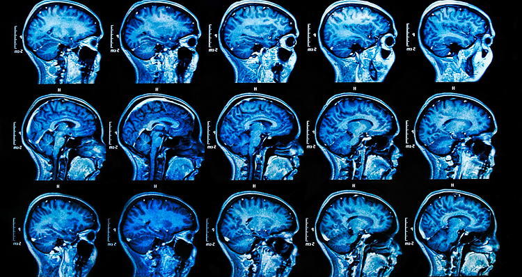
Magnetic Renausance Imaging (MRI) Scan
Nearly every part of the body can be examined in detail with an MRI scan. The MRI uses a powerful magnet, radio waves and a computer to create images of the soft tissue in your body. With that being the case tumours, traumatic injuries, multiple sclerosis, stroke, dementia and infection are visible from an MRI scan.
Advanced medicine is even recently using the MRI scan to diagnose Alzheimers, autism and Parkinson’s disease from information that is analyzed from the scan. The MRI scan is a painless and non-intrusive way to look inside your body.
Electroencephalogram (EEG)
An EEG is a test that reports the electrical activity in your brain. Your brain communicates through little electrical impulses. The impulses are active and sending signals 24 hours a day. The EEG makes a report with wavy lines. The pattern of the lines can help doctors to diagnose epilepsy, brain tumours, trauma, inflammation of the brain, stroke and even sleeping disorders. This test is painless and very safe.
Electrocardiogram (ECG)
Most times an ECC is used to diagnose heart conditions, but it is also a useful tool in diagnosing brain lesions or tumours and vascular brain injuries. Doctors can use this diagnostic tool to check for abnormal reports to help diagnose tumours early.
This test involves attaching a few special stickers around the midsection of your body and attaching wires to the stickers. The procedure is quick and painless.
Computerized Tomography (CT) Scan
A CT scan of your brain is an imaging tool that uses x-ray technology to make horizontal or axial images called “slices” of your brain. A CT scan gives doctors detailed information about your brain tissue and the structure of your brain. This scan can be done with and without contrast to allow the doctor to see even more details from your brain tissue.
During this scan, an x-ray beam moves in a circle around your head. It is noninvasive and painless. The scan is beneficial to assess head injuries, aneurysms, brain bleeds, stroke, and tumours.
Positron Emission Tomography (PET) scan
Doctors use a PET scan to see how your brain and the tissue is functioning. The test accurately reports the size, shape and function of your brain. This test requires a special dye with be added to your bloodstream. This test allows doctors to diagnose brain cancer, Alzheimer’s disease, Parkinson’s disease and to prepare for epilepsy surgery.
Using well-qualified healthcare professionals like those found at Inside Radiology is crucial for receiving the proper diagnosis and treatment. The images need to be clear, and the readings need to be accurate to be useful in your treatment plan. The tests and scans are simple for the patient but require experience and finesse from a well-trained radiology staff.
Radiologists perform many useful tests to help doctors diagnose brain disease. Most of the tests are painless, noninvasive and are relatively quick, but produce essential details of the brain to help your doctor. It is vital to use an experienced radiology clinic.